































Medical Imaging Software Development Services

Imaging Software That Speaks Radiology
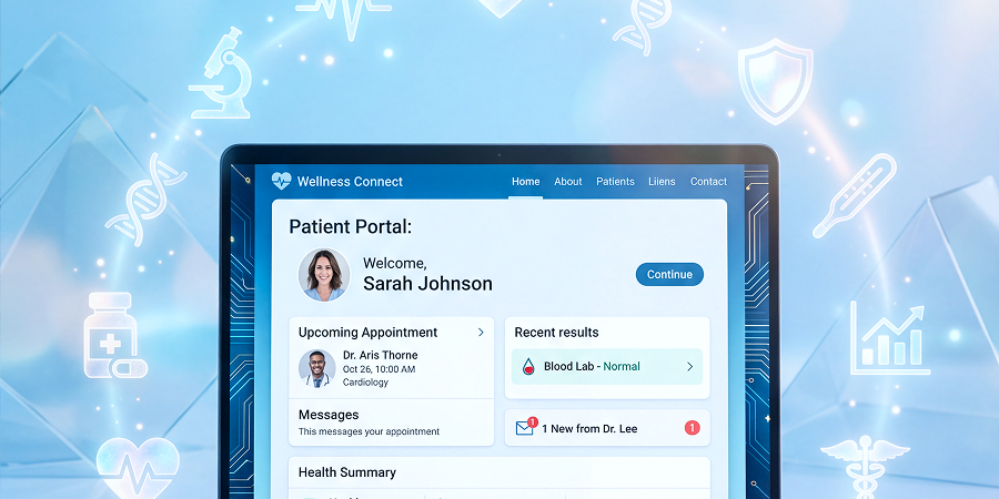
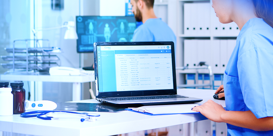
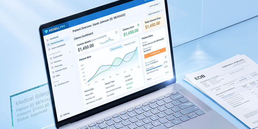
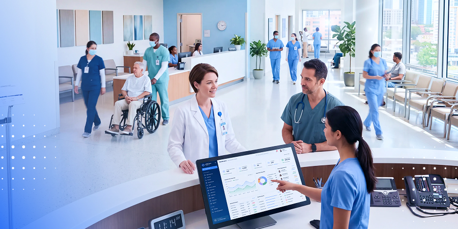
PACS, DICOM viewers, and AI-powered image analysis for CT, MRI, X-ray, ultrasound, and PET. GPU-accelerated rendering that handles large studies. FDA-ready engineering throughout.
Reliable partner
Reliable partner
Experienced team
Experienced team
Smart solutions
Smart solutions
Medical Imaging Software Development Services 1920
Medical Imaging Software Development Services 1440

Industry Leaders We Work With

Related Healthcare Services

Explore the Wider Healthcare Practice

Medical imaging software connects to the devices, records, and standards around it, so these related healthcare services cover what an imaging build depends on.

“The average radiologist reading CT or MRI must interpret one image every 3 to 4 seconds across an 8-hour day.” — National Library of Medicine

That is the pressure medical imaging software exists to relieve: a viewer that renders instantly and AI that flags findings give the radiologist back the seconds that volume has taken away.
What We Build

Medical Imaging Solutions We Build

Medical imaging software development services across the full range below, from a single DICOM viewer to a full PACS or AI analysis tool.
98%

98%

Client Retention Rate
300+

300+

Successful Projects

PACS development and integration

Custom PACS development, or integration with an existing PACS, for image storage, retrieval, and archiving.

DICOM viewers

Zero-footprint web viewers that need no install, plus desktop and mobile viewers, built to render large studies fast.

AI image analysis

Detection, segmentation, and CADx (computer-aided diagnosis) that analyze medical images inside the reading workflow.

RIS integration

Radiology information system integration for scheduling, worklist, and reporting in the radiology workflow.

3D reconstruction

3D reconstruction, MPR, and volume rendering, including 3D medical imaging software that turns a series into 3D medical images.

Image annotation

Annotation and labeling tools for AI training and for the radiologist’s read.

Teleradiology platforms

Teleradiology software for remote reading across sites.

VNA

Vendor-neutral archive (VNA) development for storage independent of any one PACS vendor.

Imaging apps

Medical imaging mobile app development services and medical imaging web development services, including zero-footprint web viewers.

Custom medical imaging

Custom medical image analysis software, and custom development for a workflow a packaged product does not fit.


Modalities and Standards

Imaging Modalities and Standards

Deep support for every modality and the standards that move images between systems is what separates an imaging engineer from a generalist, across a wide range of medical specialties.
Modalities
DICOM
DICOMweb
File formats

Every scan type

CT, MRI, X-ray, ultrasound, and PET, plus mammography, digital pathology (whole-slide imaging), and OCT software for ophthalmic imaging.
  • All major imaging modalities, from CT to digital pathology.
  • The imaging devices, medical imaging devices, and imaging hardware that produce them.

The standard

The DICOM standard, in depth: parsing, storage, and networking.
  • Digital Imaging and Communications in Medicine, the standard for medical images.
  • Both image and metadata carried between imaging systems.

Web-native exchange

Modern web integration through DICOMweb, so imaging works over standard web protocols.
  • WADO-RS for retrieval, QIDO-RS for query, STOW-RS for storage.
  • HL7 and FHIR ImagingStudy, plus IHE profiles. See our HL7 integration services.

DICOM and NIfTI

The formats clinical and research imaging actually use.
  • DICOM for clinical imaging across modalities.
  • NIfTI for research and neuroimaging.
Image abstract 1 368
Testimonials

What Our Customers Say

“In the case of Zoolatech, it's a very tight partnership.
The team at Zoolatech is incredibly collaborative, and we work as a team despite being thousands of miles away from each other.”
Spencer Rascoff
CEO Match Group
5/5
“Zoolatech has been a key technology partner for Pandora,
enhancing our software development and deployment capabilities. They're ambitious, supportive, fast-moving, and well-skilled, with sound ethical values.”
Erika Romsics
Contract and Vendor Manager, Pandora
erica
5/5
“The apps they’ve developed give us the opportunity to get more customers.
We’re providing more services to target big customers. We can install jobs faster and identify reduce bottlenecks, so we’re providing a better customer experience.”
Aida Youssef
Senior Director of Software Engineering, Complete Solaria
5/5
“Zoolatech has access to a deep talent pool and knows how to identify client's needs.
With the help of Zoolatech, went from a very early and incomplete prototype to the MVP release, the first production release, and the first paying customer!”
Greg Wagenhoffer
CEO, GreenVisr
5/5
“Zoolatech enabled us to build a world-class engineering team quickly and efficiently.
Zoolatech's pre-screening process and engineer training are customized for providing effective engineers that can contribute immediately to accelerating product roadmaps.”
Shariq Minhas
CTO, SVSG
5/5
“We can recommend Zoolatech
for their talent pool, attention, ability to understand our requirements, candidate screening process and constant communication.”
Chaitanya Pallapothula
SVP, Tailored Brands, Inc.
5/5
“Zoolatech’s developers quickly became an integral part of our team effort
with whom we shared daily stand up calls. Overall, Zoolatech fit well with our needs for agile development and continued to adapt as our needs evolved.”
Forrest Glick
UX Designer, Stanford University
5/5
“Working with Zoolatech has been a driving force in our business offerings.
The team utilizes it's experience and expertise meshing with our internal team creating a positive work environment. Zoolatech is by far one of the best teams to work with in the industry.”
Kris Naidu
CEO, Zeacon
Kris Naidu CEO, Zeacon
5/5
AI and ML

AI and Machine Learning for Medical Imaging

AI is the core of medical image analysis, so medical imaging AI software development gets a dedicated team. For the wider AI practice, see our work on AI in medical imaging.
Detection

Detection

Deep-learning detection of lesions, nodules, and fractures, flagging findings for the radiologist.
Segmentation

Segmentation

Segmentation of organs and tumors for measurement and treatment planning.
CADx and CADe

CADx and CADe

Computer-aided diagnosis and detection, AI software that supports the read rather than replaces it.
Classification

Classification

Image classification and quantification through medical image processing, turning pixels into measurable findings.
Worklist triage

Worklist triage

Workflow prioritization so urgent cases surface first in the worklist.
Governed by design

Governed by design

Built with MONAI, PyTorch, TensorFlow, and nnU-Net, under ISO 42001 AI governance, because AI that informs diagnosis is regulated.
Performance

The Future of Medical Imaging Software

Radiologists abandon viewers that lag on large studies, so we engineer for rendering performance, not just features. This is where high-performance medical imaging is won.
01

GPU-accelerated rendering

Fast rendering of large 500MB+ CT and MRI series, where Electron wrappers and legacy pipelines stall.
02

Zero-footprint web viewers

Browser-based viewers with no install, built on optimized libraries rather than desktop wrappers.
03

Open-source imaging stack

Cornerstone.js and OHIF for web viewers, ITK and VTK for processing, and Orthanc for PACS.
04

Progressive loading

Progressive loading of large studies, so a radiologist starts reading before the whole series arrives.

“AI reading CT scans for pulmonary embolism missed 23 cases; the attending radiologist missed 60.” — National Library of Medicine


AI does not replace the radiologist; it catches what a tired one misses. That is why detection and CADx belong in the reading workflow.
How We Build

The Medical Imaging Development Process

Imaging software is regulated, production-grade software, so our development process runs a disciplined development lifecycle from concept to validated release.
Step 1

Discovery and design

We map modalities, workflow, integrations, and regulatory pathway, then move to designing the software and its architecture before any code.
Step 2

Build and integrate

Our software developers build in sprints, integrating PACS, RIS, and EHR early, because integration is where imaging software development projects usually slip.
Step 3

Validate and release

Testing, validation, and software implementation to production, with the documentation regulated software requires. Managing overall development this way keeps development time predictable.
Integration

Integration: PACS, RIS, and EHR

Medical imaging software is only useful inside real imaging workflows, so we integrate imaging across the systems radiology runs on.

PACS, RIS, and EHR

Imaging connected across PACS, RIS, and the EHR through FHIR ImagingStudy, so images reach the patient record. See our EHR software development.

Worklist and routing

Worklist management, DICOM routing, and modality worklist (MWL), so studies reach the right radiologist automatically and nothing sits unread.

Exchange

Health information exchange and cross-site sharing, so medical information and medical data follow the patient across the imaging domain.
Our Approach

Where Zoolatech Excels in Medical Imaging

What makes a medical imaging software development company worth hiring is technical depth, and that is what we compete on, from a real imaging stack to performance on large studies.
check icon

Technical depth

A real imaging stack, from Cornerstone.js and OHIF to MONAI, ITK, VTK, and Orthanc, not a generic framework dressed up for healthcare.
check icon

Performance

Engineered for large studies and GPU rendering, the thing radiologists judge a viewer on first.
check icon

Regulated experience

Proven on FDA-regulated software with MasterControl, so the 510(k) mindset is built in.
check icon

AI expertise

Imaging-specific AI, from detection to CADx, under ISO 42001 governance.
check icon

Product development

As a medical imaging product development company, our consultants take a medtech product from concept to an FDA-ready build, not just a feature list.
check icon

Senior-heavy delivery

A senior team of software developers that has delivered 300+ projects since 2017, with 600+ engineers and 96% client satisfaction.
check icon

Engagement models

Managed delivery, team extension, or a dedicated development team, with a Miami HQ and delivery centers in Poland, Ukraine, Mexico, and Turkey.
Technology Stack

The Medical Imaging Stack

The stack decides how fast a viewer renders, how cleanly it integrates, and how well AI performs.
Cornerstone.js
Cornerstone.js
OHIF
OHIF
MONAI
MONAI
ITK
ITK
VTK
VTK
Orthanc
Orthanc
AWS
AWS
PyTorch
PyTorch
TensorFlow
TensorFlow
Azure
Azure
Python
Python
React
React
SQL
SQL
and other
Why Choose Us

Why Businesses Trust Us

logo
At Zoolatech, we create engineering teams for industry leaders across the US and Europe — teams that move fast, think big, and deliver strong impact.
96%
Client Satisfaction
300+
Successful Projects
2017
Year Founded
98%
Retention Rate
team sport photo
At Zoolatech, we create engineering teams for industry leaders across the US and Europe — teams that move fast, think big, and deliver strong impact.
Engineering Excellence. Every Time.
main award png (1)
At Zoolatech, we create engineering teams for industry leaders across the US and Europe — teams that move fast, think big, and deliver strong impact.
team sport photo
600+
Employees
Headquarters
USA
Development Centers
PL
UA
MX
TR

Plan Your Software Implementation

Tell us your modalities, whether you need a viewer, PACS, or AI analysis, and your regulatory pathway. We'll scope an approach.

Questions You May Have

What is medical imaging software development?

Medical imaging software development is the engineering of applications that acquire, store, display, and analyze medical images: CT, MRI, X-ray, ultrasound, and PET scans. It includes PACS, DICOM viewers, and AI tools for detection and segmentation. Because it processes diagnostic data, medical imaging software follows the DICOM standard, integrates with RIS and EHR systems, and may require FDA clearance as Software as a Medical Device when used for diagnosis.

What is DICOM and why does medical imaging software use it?

DICOM (Digital Imaging and Communications in Medicine) is the international standard for storing, transmitting, and displaying medical images and their metadata. Every modality, from CT to MRI to X-ray, produces DICOM files containing both the image and patient and acquisition data. Medical imaging software uses DICOM so images move reliably between scanners, PACS, viewers, and other systems. Modern integrations also use DICOMweb (WADO-RS, QIDO-RS, STOW-RS) to exchange images over standard web protocols.

How is AI used in medical image analysis?

AI in medical image analysis automates and assists the analysis of medical images that once required manual reading. Deep-learning models detect abnormalities such as lung nodules, fractures, or lesions; segment organs and tumors for measurement; classify findings; and triage worklists so urgent cases surface first. These are built with frameworks like MONAI, PyTorch, and TensorFlow. When AI output informs diagnosis, the software is regulated as a medical device and must follow an FDA clearance pathway.

Why does medical image processing software struggle with large studies?

Many DICOM viewers lag on large CT or MRI series, often hundreds of megabytes, because they rely on architectures not optimized for big medical imaging data, such as general-purpose web wrappers or older rendering pipelines. Performance improves with GPU-accelerated rendering, progressive image loading, and zero-footprint web viewers built on optimized libraries like Cornerstone.js and OHIF. Radiologists abandon tools that lag, so rendering performance is a core engineering requirement, not a finishing touch.

What does medical imaging software integrate with?

Medical imaging software integrates with PACS for image storage and retrieval, RIS for radiology workflow and scheduling, and EHR systems for the patient record, using DICOM, DICOMweb, and HL7 or FHIR ImagingStudy resources. It also connects to modality worklists, vendor-neutral archives (VNA), and health information exchanges. AI analysis tools integrate into the reading workflow so results appear alongside images in the radiologist’s viewer rather than in a separate system.

How is medical imaging software used across different medical specialties?

Medical imaging software helps across a wide range of medical specialties: radiology, cardiology, oncology, ophthalmology (where OCT software reads retinal scans), and pathology. Each medical practice has its own imaging needs and modalities, so imaging technologies and imaging systems are configured to how that specialty reads, measures, and reports. The future of medical imaging software is more AI assistance and better access to medical images at the point of care.

Does medical imaging software need FDA clearance?

It depends on intended use. Software that only displays or stores images for non-diagnostic purposes is generally not regulated. Software that analyzes images to detect, diagnose, or measure disease qualifies as Software as a Medical Device (SaMD) and typically requires FDA 510(k) clearance. AI diagnostic tools almost always require clearance. Classification should be determined early, because it shapes the development lifecycle under IEC 62304 and the validation and documentation required.

How much does medical imaging software development cost?

Medical imaging software development cost depends on the type of software, a DICOM viewer, a full PACS, or an AI analysis tool, the modalities supported, integration scope, and whether FDA clearance is required. An AI diagnostic tool requiring 510(k) clearance and clinical validation costs substantially more than a viewer. A precise estimate follows a discovery phase that maps your modalities, workflow, integrations, and regulatory pathway.